Project 20 interviews
«Professionals speak about their daily work and their missions»
Interview with Dr. Olfa Chouchane Mlik – Head of the Pathological Anatomy Department of the National Health Laboratory (LNS) in Dudelange
Pathological anatomy is a medical discipline practiced by specialized doctors called anatomopathologists or pathologists.
Pathological anatomy is based on the analysis of tissues or cells taken by biopsy, cytopuncture or during a surgical operation.
The purpose of the examination of these samples is to determine the nature of the lesion, its benign or malignant status, its grade and its stage in the case of cancer.
Histopathological analysis of breast cancer has long been limited to the classical morphological study. It now benefits from the contribution of complementary techniques such as immunohistochemistry and molecular biology.
The results obtained by these different technical procedures make it possible to characterize diagnosed breast cancers according to international recommendations. The pathologist’s final diagnosis provides vital information that conditions the therapeutic approach to breast cancer and its follow-up.
Three types of samples are received in the context of breast pathology
In the pathology laboratory, three types of samples are received in the context of breast pathology:
1. Cytopuncture: fine needle puncture of a pathological axillary lymph node or a clinically or radiologically identified breast area to collect cells.
2. Biopsy: removal of a small fragment from an abnormal area of the breast or in the presence of microcalcifications.
3. Surgical specimen obtained by surgical removal.
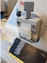
Technical steps in the management of a breast biopsy
The analysis of these samples begins with a macroscopic examination. An incompressible fixation time of 6 hours must be respected to guarantee optimal analysis. A description of the lesions and their dimensions will be carried out, followed by a sampling of the surgical specimen. The tissue samples will be placed in a plastic box (a cassette), which will be placed for a few hours in a tissue impregnation machine. This is to prepare the sample for paraffin embedding. To do this, it is essential to dehydrate the tissue sample to be analyzed so that it becomes miscible in the paraffin.
Once this dehydration process is complete, the cassette is removed from the machine. This is followed by inclusion in very hot paraffin, which is frozen at low temperature to recover a hard block. This can be stored for years. This tissue block will then be cut by the technicians into ultra-fine sections of 3 to 5 microns. These sections will be spread out on a glass slide. They are then stained (hematoxylin and eosin stain) to make the tissue visible under the light microscope.
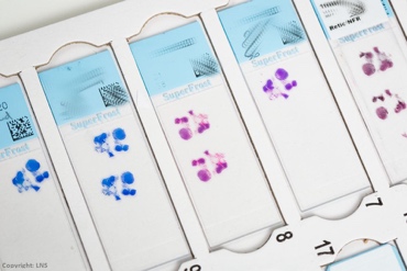
The pathologist will examine the resulting section(s) under the microscope to identify the breast lesion and classify it (inflammatory, degenerative or tumourous).
The immunohistochemical study detects the presence or absence of markers expressed by the tumour cells. It allows the quantification of hormone receptors and other markers that allow the prediction of sensitivity or not to a treatment. This step is essential for the subsequent therapeutic management of patients.
In some situations, molecular biology techniques will be used to identify abnormalities in the cell genome such as mutations or gene amplifications, thus allowing the classification of the cancer to be refined or providing prognostic information.
Considering the technical constraints necessary to achieve a complete analysis, it is understandable that results cannot be provided in a few hours. Biopsies are processed as a priority. It takes 4 to 5 working days for the analysis and the complete report (including the analysis of biomarkers). This will help the specialists to choose the appropriate treatment after discussion at multidisciplinary consultation meetings.
The anatomopathological diagnosis of cancers is constantly improving. Our vision,” concludes Dr. Chouchane, “is to provide a service of excellence based on the application of international standards. Dr. Chouchane also encourages patients to follow up on their invitations for the mammography carried out under the Luxembourg national program. Don’t hesitate to consult your GP,” she says. It is even more difficult to control the disease if it is discovered at an advanced stage. That is why regular consultations are very important, precisely so that any abnormality can be detected at the earliest possible stage.
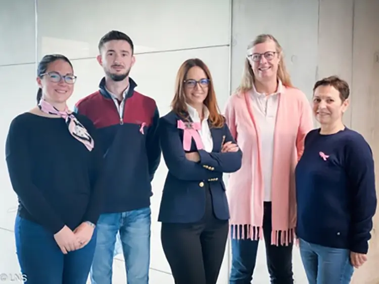
Team of gynaecopathologists from the LNS Pathology Department
From left to right: Dr Alexandra Oniga, Dr Flaviu Crisan, Dr Olfa Chouchane Mlik, Dr Anne Janssen, Dr Alena Badsi
Here is a link to a video on youtube, filmed in the lab, which illustrates the technical details reported in the interview:
Link: https://www.youtube.com/watch?v=CkaQgY4Y_P0&t=131s
The interview was conducted by Ms. Françoise Hetto-Gaasch, member of the committee of Europa Donna Luxembourg in May 2022.
Europa Donna Luxembourg Asbl
1b rue Thomas Edison L-1445 Strassen
Tél. : 621 47 83 94
E-mail : europadonna@pt.lu