Project 20 interviews
«Professionals speak about their daily work and their missions»
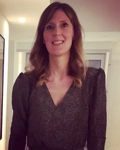
Interview with radiologist Dr. Audrey Mawet: her profession, her role, her commitment, her responsibility
Dr Mawet, who works at the CHdN, is a radiologist specialized in breast cancer diagnosis.
She explains what her work involves.
As a senologist, Dr Mawet uses her expertise in the following situations:
- asymptomatic patients, over 40, are prescribed screenings by their gynaecologist. Patients under 40 may also be included, where there is a history of breast cancer in the family. For example, if the patient has a close family member who was diagnosed with cancer under the age of 40.
- patients, aged between 50 and 70, invited to take part in the national mammogram screening program
- urgent patients with symptoms (such as a lump, discharge from the nipple….) referred directly by their gynaecologist
It should be noted that men present with different types of symptoms such as sore swellings, lumps, or a skin retraction. In these cases, biopsies/samples are usually sent immediately to a lab to be analyzed, and a mammogram and ultrasound are done.
A mammogram looks at the inner structure of the breast, and in this way can detect any abnormality of the breast. A 2-D scan is provided in black/grey and white:
- white signifies mostly fibrous-glandular tissue, and abnormalities such as a malignancy or a cyst
- black/grey represents breast fat-tissue
By analyzing the scans, the radiologist is able to recognize the different existing abnormalities in the breast.
In the case of breast cancer, the terms used are spongy opacity, architectural distortion, or suspect microcalcifications.
In some cases, it is important to include an ultrasound to use more precision in mammograms. And often it is used for very young patients, or for women with dense breasts.
The radiologist has huge responsibility when examining such scans.
If an abnormality is detected, the radiologist suggests that the patient has a biopsy. This shows whether or not the abnormality is benign.
A biopsy involves taking a sample of tissue of the lesion. This sample will be sent to the national laboratory of health, in order to be examined and in order to define the characteristics of the lesion.
The patient will have to wait 7 to 10 days to get the result. Perhaps this might seem too long, but it allows the patient to come to terms with a possible serious outcome.
If the radiologist is convinced that it is a benign lesion, she can suggest another type of biopsy, such as cytopunction by which some cells of the breast lesion are sucked up by a fine needle. Of course, this diagnosis is less precise but sufficient for the follow up of the lesion.
During these different technical procedures, for which radiologists are responsible, they need to be empathic and sensitive in order to explain all the details clearly to their patient. This is a relationship based on trust.
Finally, the results are communicated to the patient by the gynecologist or general practitioner.
If the patient has breast cancer, the radiologist will then only see her/him for a yearly screening.
If chemotherapy has to be done before surgery, the radiologists will see their patient before surgery, in order to see if chemotherapy has had a successful impact on the tumor by diminishing its size.
The radiologist also has to regularly check certain benign lesions, according to established guidelines.
By the end of the consultation, the radiologist has to do a classification according to ACR (American College of Radiology), which will define further treatment.
In addition to a mammogram and an ultrasound, an MRI may be carried out for patients who present with a genetic mutation (BRCA type or others), or in cases of certain types of breast cancer.
Dr Mawet, the radiologist is happy to announce that in 2 years a new technique of screening will be available to hospitals in Luxembourg. This new technique, called tomosynthesis, is able to provide 3-D screening, therefore being much more precise. This machine will have a higher rate of detection, leading to more precise diagnosis.

For Dr Mawet mammograms are an essential part of a process for early detection.
If a cancer is discovered early, rates of survival increase enormously.
These days treatment has evolved, and is more targeted.
Dr Mawet encourages all patients to ask questions, so that they are fully aware of the tests that are needed, the discomfort that might be felt during mammograms, and their fears and apprehension of X-rays.
Thank you so much Doctor Mawet for your dedication, and the time spent giving us this vital information.
The interview was conducted by Ms. Françoise Hetto-Gaasch, member of the committee of Europa Donna Luxembourg in May 2022.
Europa Donna Luxembourg Asbl
1b rue Thomas Edison L-1445 Strassen
Tél. : 621 47 83 94
E-mail : europadonna@pt.lu
